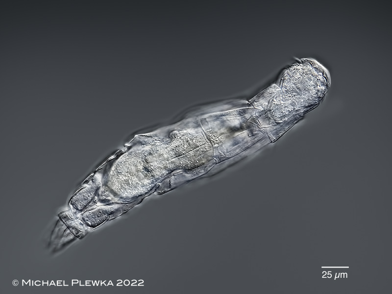
Dicranophorus cambari: (dorso)ventral view; focus plane on the ventral ciliary field of the head.
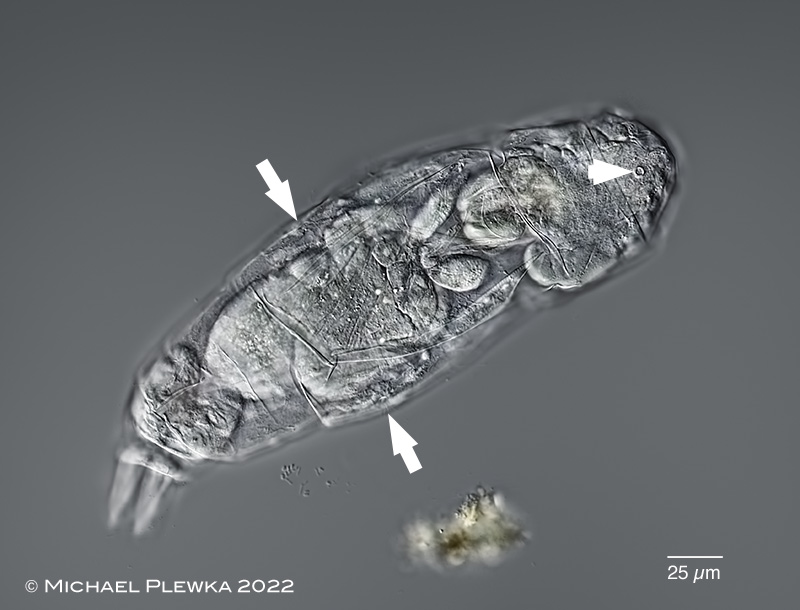
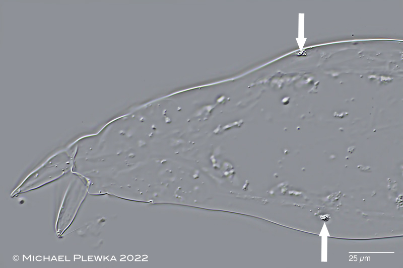
Dicranophorus cambari: dorsal view. The arowhead points to the opening of the dorsal antenna, whereas the arrows mark the locations of the lateral antennas.
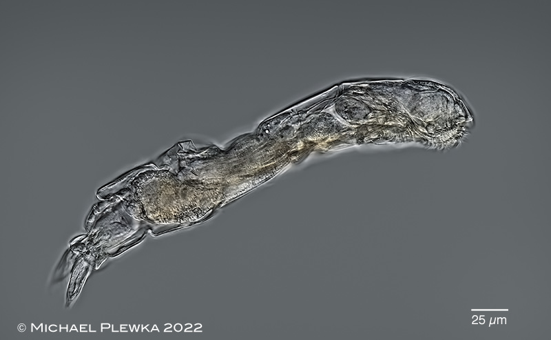
Dicranophorus cambari: swimming, lateral view.
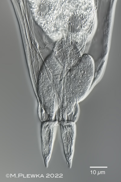
Dicranophorus cambari: foot with toes and distinct tips. Also visible are the footglands. Slightly compressed specimen.
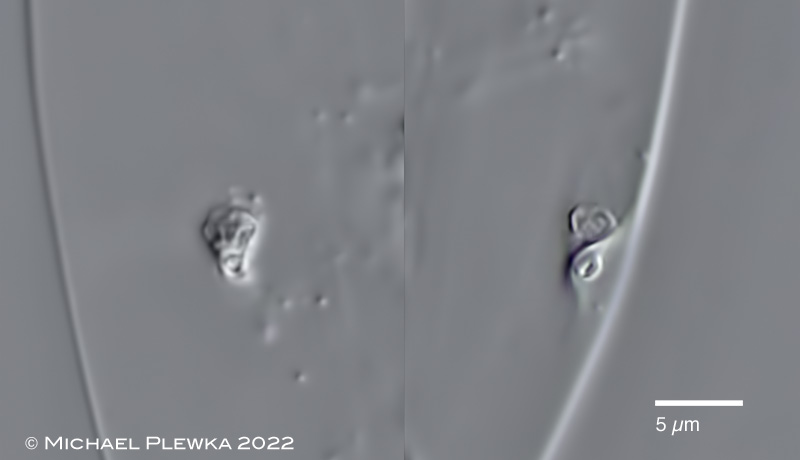
Dicranophorus cambari: upper image: the arrows point to the openings of the lateral antennas in this specimen which is semimacerated with SDS. Lower image: combination of two crops of the upper image.
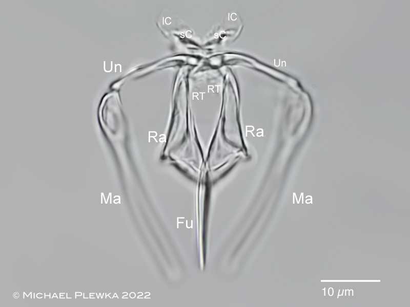
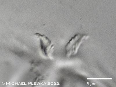
Dicranophorus cambari: the forcipate trophi of this morphotype are characterized by the special shape of the epipharyngeal structures (lC; sC) that differ for example from the Dicranophorus hauerianus-morphotype found in France. Fu: fulcrum, Ma: manubrium; Ra: ramus; RT: rami teeth (in this morphotype: needle-shaped); lC: large epipharyngeal comb; sC: small epipharyngeal comb; Un: unci.
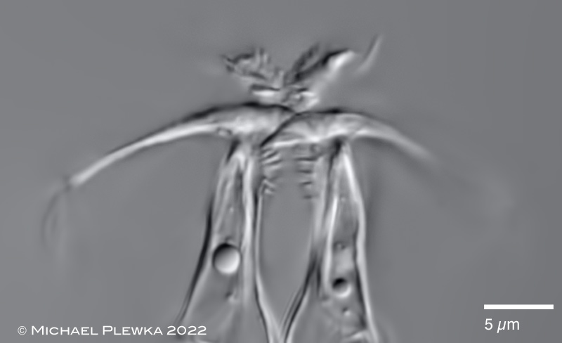
Dicranophorus cambari: distal part the forcipate trophi of another specimen. The "bubbles" are lipid droplets from the stomach.
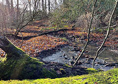
Habitat: detritus/ sediment in lotic water. In contrast to the observations of De Smet and Verolet (2016) (epibiotic on Gammarus pulex) the specimens presented here were found free living.